Annotate and visualize plasmids in one click
Two isolates can look unrelated and still share the same resistance plasmid. Plasmids spread by horizontal transfer, moving between bacteria and sometimes across species. A single plasmid can hold several resistance genes at once, so one transfer passes all of them to the new host.
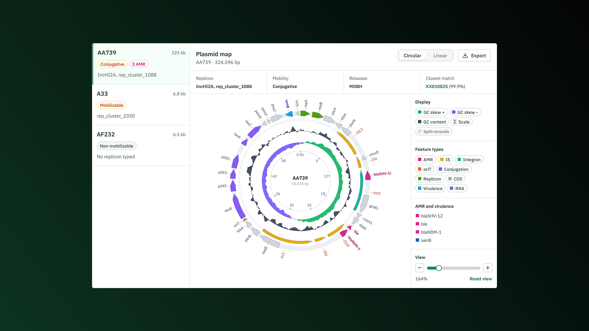
Solu identifies a plasmid's AMR and virulence genes, integrons, IS elements, replication and transfer machinery, and all other coding sequences. It then draws them on a clear map you can explore, adjust, and export.
How it works
With Solu, you can run a full plasmid analysis with one click.
- Choose a sample. Solu lists every plasmid reconstructed from the assembly, with its size, mobility and AMR gene count.
- Run the analysis. Pick a plasmid to start. Its AMR and virulence genes, integrons, IS elements (transposons), and other coding sequences are annotated, then the map is built.
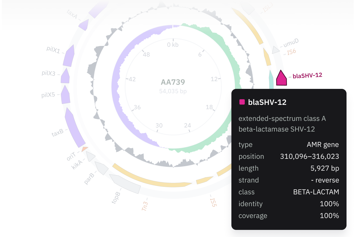
- Explore the map. Hover any feature to read its position, length, strand and other details.
- Adjust the view. Show or hide any feature type or GC track, switch between circular and linear, and zoom in on any region.
- Export the result. The map can be exported as an image, the combined annotation as a GFF3 file, and the plasmid sequence as FASTA.
What you can see
- Annotation. AMR and virulence genes, integrons, IS elements, replication and transfer machinery, all other coding sequences, plus tRNA, rRNA, tmRNA and ncRNA.
- Gene position. Where each gene sits on the plasmid: coordinates, strand, and what it neighbors.
- Mobility. Replicon family, relaxase type, mate-pair formation type, and predicted mobility: conjugative, mobilizable or non-mobilizable.
- GC content and skew. Both plotted around the plasmid. Composition shifts can flag acquired DNA, and the skew inversion marks the replication origin.
- Closest public match. The most similar plasmid in public databases, with a similarity score.
How the annotation is produced
The annotation runs on established open tools, and the result is one view that shows not just what a plasmid carries, but where each part is located.
- MOB-suite for plasmid reconstruction and typing
- Bakta for genes and other features
- ISEScan for insertion sequences
- IntegronFinder for integrons
AMR and virulence genes come from Solu's sample-level AMRFinderPlus and VFDB results, mapped onto the plasmid.
If you're interested in reading more about our methodology, check out our methodology guide.
Try it today
Plasmid annotation and visualization is available in Solu today. Log in, open any sample's plasmids tab, and build your first map. If you don't have an account yet, start a free trial and try Solu on your own data.
.png)
.png)